- The anterior fat pad of the elbow can be visualized normally as a
thin radiolucent line just anterior to the coranoid fossa (anterior border of
the distal humerus). However, when the elbow joint becomes distended (i.e.,hemarthrosis
secondary to fracture within the joint space), the anterior fat pad is
displaced further anteriorly and superiorly to form an anterior "Sail
Sign," or more prominent lucency.
- The posterior fat pad lies over the olecranon fossa and
normally is not visible because
the olecranon fossa is much deeper (more concave) than the coranoid fossa.
Visualization of the posterior fat pad (even as only a thin radiolucentline on
the lateral view) indicates marked distention of the joint capsule, due to
hemarthrosis from an intra-articular fracture and is therefore always
pathologic.
- The anterior humeral line is drawn along the anterior surface of
the distal humeruson a true lateral. Normally this line should intersect the
middle third of the capitellum. If there is a supracondylar fracture with
posterior displacement of the distal segment, the anterior line will either
intersect the anterior third of the capitellum or does not intersect the
capitellum at all.
- The radiocapitellarline is drawn along the central axis of the
radius on the lateral view. Normally, this line should intersect the center of
the capitellum (in all views). If the line does not transect the middle of the
capitellum, either the radial head is dislocated or there is a fracture
through the radial neck region.

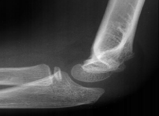
On this true lateral:
- There are both an anterior and posterior fat pads.
- The anterior humeral line intersects the anterior third of the capitellum
rather than the middle third.